File:Hip replacement Image 3684-PH.jpg

File originale (5 556 × 4 468 pixel, dimensione del file: 602 KB, tipo MIME: image/jpeg)
| Questo file e la sua pagina di descrizione (discussione · modifica) si trovano su Wikimedia Commons (?) |
| Descrizione |
Čeština: Totální endoprotéza kyčelního kloubu
Deutsch: Röntgenaufnahme einer Hüfte. Das rechte Hüftgelenk des Patienten (im Bild links) wurde durch eine Prothese ersetzt.
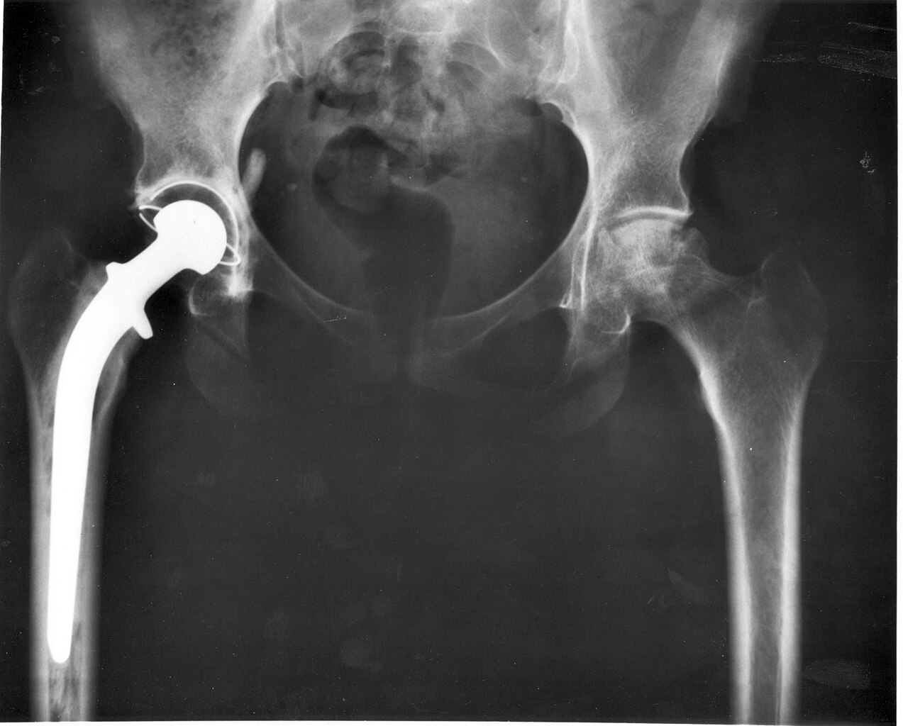
English: An A-P X-ray of a pelvis showing a total hip joint replacement. The right hip joint (on the left in the photograph) has been replaced. A metal prostheses is cemented in the top of the right femur and the head of the femur has been replaced by the rounded head of the prosthesis. A white plastic cup is cemented into the acetabulum to complete the two surfaces of the artificial "ball and socket" joint.
Although not the case here, hip prostheses can also be made of a ceramic material which rarely wears out during the patient's lifetime. During the operation, a bonding cement is used to fix the metal prostheses into the shaft of the femur and the plastic cup to the acetabulum (socket in the hip bone). One of the leading reasons for hip replacement is osteoarthritis of the hip joint in which virtually all of the cartilage around the top of the femur bone deteriorates due to wear over time, leaving a grinding bone-on-bone situation with the bone surfaces becoming roughened leading to pain and stiffness. Narrowing of joint space (the space between the acetabulum and the head of the femur) is also a feature of osteoarthritis. There may be other changes which are not entirely clear on this A-P X-ray, which probably should be reported together with lateral X-rays of the hips, or with modern computerised imaging techniques. Keywords: total hip replacement, prosthesis, osteoarthritis, X-ray.Français : Remplacement total de l'articulation de la hanche.
La hanche droite de ce patient (à gauche sur la photo) a été remplacée par une articulation artificielle. L'articulation artificielle mime les formes naturelles, la tête du fémur étant remplacée par une tête métallique insérée dans la moële de l'os, et s'insérant dans une cupule en plastique blanc insérée dans le bassin. L'une des raisons les plus fréquentes de remplacement de l'articulation de la hanche est souvent l'arthrose.
Tiếng Việt: Ảnh chụp X quang của một xương chậu và hông. Khớp hông bên phải của bệnh nhân (trong ảnh là bên trái) đã được thay thế bởi một bộ phận giả. |
||||||
| Fonte | from NIH Here. Credit: NIADDK, 9AO4 (Connie Raab-contact); NIH. | ||||||
| Autore | X-ray Image ID: 3684. Photographer: Unknown. | ||||||
| Licenza (Riusare questo file) |
|
||||||

|

Questa immagine è stata selezionata come Immagine del giorno in data 12 agosto 2006. La didascalia era la seguente: Italiano: Immagine ai raggi X di un cinto pelvico ricostruito artificialmente attraverso una struttura metallica incassata in una cavità di materiale plastico Altre lingue:
Cymraeg: Delwedd Peledr-X yn dangos gwregys pelfig artiffisial. English: The patient’s right hip joint replaced by a metal head and a plastic cup. Español: Cadera derecha del paciente reemplazada por una cabeza de metal y una semiesfera de plástico. Galego: Na imaxe de raios X vese como a cabeza do femúr foi substituída na cadeira dereita dun paciente por unha peza de metal que encaixa nunha semi-esfera de plástico Italiano: Immagine ai raggi X di un cinto pelvico ricostruito artificialmente attraverso una struttura metallica incassata in una cavità di materiale plastico Magyar: Jobb oldali csípőprotézis fém fejjel és műanyag borítással. Norsk bokmål: En pasients øvre hofteledd er blitt erstatta med en erstatning av metall og plast. Norsk nynorsk: Det høgre hofteleddet til pasienten er erstatta av ei indre protese i metall og plast. Polski: Zdjęcie rentgenowskie pacjenta ze sztucznym prawym stawem biodrowym. Português: A articulação da perna direita no quadril de um paciente, substituída por uma cabeça de metal e um encaixe plástico. Svenska: Rötgenbild av en höftled som ersatts med en protes. Русский: Рентгенограмма металлического протеза правого тазобедренного сустава с пластмассовой вертлужной чашкой 日本語: 人口股関節置換術を施された右股関節(向かって左側)のレントゲン写真 中文: 由金属股骨头和塑料骨臼替换的右髋关节X光照片。 العربية : ورك استعاضي من المعدن و كأس بلاستيكي. |
This image was selected as picture of the day on Vietnamese Wikipedia.
|
Cronologia del file
Fare clic su un gruppo data/ora per vedere il file come si presentava nel momento indicato.
| Data/Ora | Miniatura | Dimensioni | Utente | Commento | |
|---|---|---|---|---|---|
| attuale | 19:32, 19 mag 2006 | | 5 556 × 4 468 (602 KB) | Gorgo | high-res version |
| 12:01, 17 mag 2006 |  | 746 × 600 (34 KB) | Teveten | from en.wiki http://en.wikipedia.org/wiki/Image:Hip_replacement_Image_3684-PH.jpg {{PD-USGov-HHS-NIH}} |
Pagine che usano questo file
Le seguenti 3 pagine usano questo file:
Utilizzo globale del file
Anche i seguenti wiki usano questo file:
- Usato nelle seguenti pagine di af.wikipedia.org:
- Usato nelle seguenti pagine di ar.wikipedia.org:
- Usato nelle seguenti pagine di as.wikipedia.org:
- Usato nelle seguenti pagine di ba.wikipedia.org:
- Usato nelle seguenti pagine di bn.wikipedia.org:
- Usato nelle seguenti pagine di ca.wikipedia.org:
- Usato nelle seguenti pagine di da.wikipedia.org:
- Usato nelle seguenti pagine di de.wikipedia.org:
- Usato nelle seguenti pagine di en.wikipedia.org:
- Usato nelle seguenti pagine di en.wiktionary.org:
- Usato nelle seguenti pagine di es.wikipedia.org:
- Usato nelle seguenti pagine di eu.wikipedia.org:
- Usato nelle seguenti pagine di fa.wikipedia.org:
- Usato nelle seguenti pagine di fi.wikipedia.org:
- Usato nelle seguenti pagine di fr.wikipedia.org:
- Usato nelle seguenti pagine di fr.wiktionary.org:
- Usato nelle seguenti pagine di gl.wikipedia.org:
- Usato nelle seguenti pagine di he.wikipedia.org:
- Usato nelle seguenti pagine di hr.wikipedia.org:
- Usato nelle seguenti pagine di hu.wikipedia.org:
- Usato nelle seguenti pagine di hy.wikipedia.org:
Visualizza l'utilizzo globale di questo file.